
A Clinician’s Guide to the 8 Main Types of Chronic Wounds in 2025
A wound that doesn't heal is more than just a break in the skin; it's a complex clinical challenge that signals a disruption in the body's natural repair mechanisms. Chronic wounds, formally defined as those that fail to proceed through an orderly and timely reparative process, affect millions of people and represent a significant burden on healthcare systems.
Far from being a single entity, these persistent injuries arise from a multitude of causes, each requiring a distinct diagnostic and therapeutic strategy. Differentiating between the various types of chronic wounds is the foundational step toward effective management and successful outcomes. An accurate diagnosis prevents treatment delays, avoids therapies that could be harmful, and paves the way for advanced interventions that can accelerate healing. Most importantly, it helps prevent devastating complications like severe infection or amputation.
This guide provides a comprehensive roundup of the primary categories of chronic wounds, from common pressure injuries to complex arterial ulcers. We will explore the defining characteristics of each, offering clinicians, caregivers, and patients a clear roadmap for:
- Identification: Pinpointing the specific wound type based on clinical features.
- Management: Understanding common treatment approaches and when to seek specialty care.
- Prevention: Implementing targeted strategies to reduce the risk of occurrence and recurrence.
By breaking down the key types of chronic wounds, this article offers actionable insights to improve diagnostic accuracy, streamline care, and ultimately support the healing journey.
1. Pressure Injuries (Pressure Ulcers/Bedsores)
Pressure injuries, often called pressure ulcers or bedsores, are localized damage to the skin and underlying soft tissue. They typically occur over bony prominences like the sacrum, heels, hips, and elbows. This type of chronic wound develops from intense and prolonged pressure, or pressure combined with shear or friction, which cuts off blood supply to the tissue, leading to cell death.
These injuries are a significant concern for individuals with limited mobility, such as those who are bed-bound, wheelchair users, or patients in long-term care facilities. The severity is categorized into stages, ranging from Stage 1 (non-blanchable redness on intact skin) to Stage 4 (full-thickness tissue loss with exposed bone, tendon, or muscle), and also includes unstageable and deep tissue pressure injuries.

Key Causes and Risk Factors
The primary cause is sustained pressure that exceeds capillary blood pressure, obstructing blood flow. Key risk factors include immobility, sensory impairment (like in spinal cord injuries), malnutrition, dehydration, and medical conditions affecting blood flow such as diabetes and vascular disease. Excessive moisture from incontinence can also weaken the skin, making it more susceptible to breakdown. Preventing skin breakdown is paramount in avoiding pressure injuries. A practical guide to effective perineal care highlights essential techniques for patient care.
Diagnosis and Treatment
Diagnosis begins with a thorough skin assessment, often using validated risk scales like the Braden or Norton scales to identify at-risk patients. Clinicians look for signs of tissue damage, such as changes in skin color, temperature, or firmness. Early detection is critical, and advanced diagnostic tools can play a role. Point-of-care fluorescence imaging, for example, helps clinicians detect and locate elevated bacterial presence in and around the wound, which can guide treatment and prevent infection. To explore how this technology aids in managing complex wounds, you can learn more about point-of-care wound imaging.
Treatment focuses on removing pressure from the affected area, keeping the wound clean and dressed, managing pain, and ensuring proper nutrition. Key strategies include:
- Regular Repositioning: Turning and repositioning patients at least every two hours.
- Support Surfaces: Using pressure-reducing mattresses, cushions, and other devices.
- Wound Care: Selecting appropriate dressings to maintain a moist healing environment and debriding necrotic tissue when necessary.
- Nutrition: Ensuring adequate intake of protein, calories, vitamins, and minerals to support tissue repair.
Referral to a specialty wound care clinic is recommended for non-healing Stage 2 wounds or any Stage 3 or 4 injuries, as they often require advanced therapies like debridement, specialized dressings, or negative pressure wound therapy.
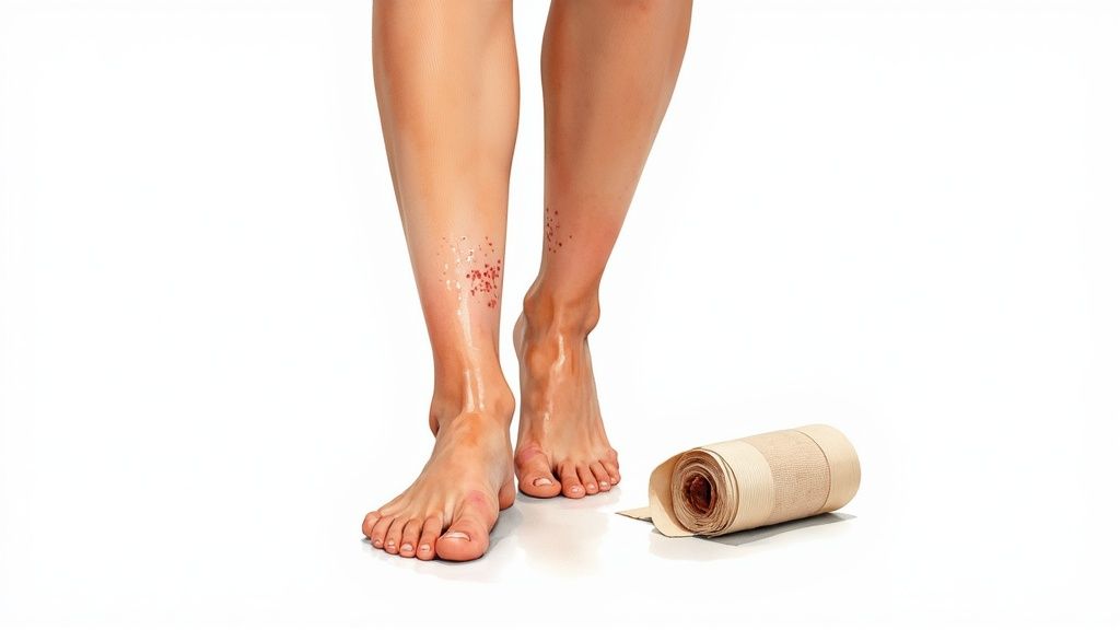
2. Venous Leg Ulcers
Venous leg ulcers (VLUs) are the most common type of lower extremity ulcer, accounting for approximately 70% of all leg ulcers. They arise from chronic venous insufficiency (CVI), a condition where the veins in the legs, particularly the valves, fail to efficiently return blood to the heart. This leads to a buildup of pressure in the lower leg veins (venous hypertension), which causes fluid to leak into the surrounding tissues, leading to edema, inflammation, and eventual skin breakdown.
These wounds typically develop on the medial (inner) side of the lower leg, often just above the ankle, an area known as the "gaiter region." They are often shallow, have irregular borders, and may be accompanied by a significant amount of exudate (fluid drainage). The surrounding skin frequently shows signs of venous disease, such as varicose veins, hemosiderin staining (a brownish discoloration), and lipodermatosclerosis (hardening and tightening of the skin).
Key Causes and Risk Factors
The root cause of VLUs is sustained venous hypertension from CVI. Key risk factors that contribute to this condition include a history of deep vein thrombosis (DVT), varicose veins, obesity, multiple pregnancies, and occupations that require prolonged standing. Other contributing factors are advanced age, limited mobility, a family history of venous disease, and previous trauma to the leg.
Diagnosis and Treatment
Diagnosis is primarily clinical, based on the characteristic appearance and location of the ulcer along with a patient history consistent with CVI. An Ankle-Brachial Index (ABI) test is crucial to rule out significant arterial disease before initiating compression therapy, which is contraindicated in cases of severe arterial insufficiency. Doppler ultrasound studies can confirm venous reflux and identify incompetent veins.
The cornerstone of VLU treatment is managing the underlying venous hypertension. Key strategies include:
- Compression Therapy: Applying sustained, graduated compression (usually 30-40 mmHg) via multi-layer bandages or stockings is the gold standard. This reduces edema and supports venous return.
- Leg Elevation: Regularly elevating the legs above the level of the heart helps reduce swelling and venous pressure.
- Wound Care: This involves cleaning the wound, managing exudate with appropriate absorptive dressings to maintain a moist environment, and debriding any non-viable tissue.
- Exercise: Encouraging calf muscle pump exercises, such as walking or ankle flexions, improves circulation.
For complex, non-healing ulcers, a referral to a specialty wound care clinic is necessary. They may consider advanced treatments, including advanced biologic skin substitutes to promote healing. Explore how a dual-layer allograft can aid in healing venous leg ulcers. In some cases, surgical interventions like vein ablation may be required to correct the underlying venous reflux.
3. Diabetic Foot Ulcers
Diabetic foot ulcers (DFUs) are a serious complication of diabetes, developing from a combination of peripheral neuropathy (nerve damage causing loss of sensation), peripheral arterial disease (poor circulation), and impaired healing. These open sores, typically located on the bottom of the foot, occur in approximately 15% of individuals with diabetes and are a leading cause of non-traumatic lower limb amputations. Even minor cuts or blisters can progress into severe ulcers because the patient may not feel the initial injury.
Key Causes and Risk Factors
The primary contributors are neuropathy, which diminishes the protective sensation of pain, and peripheral arterial disease, which reduces blood flow needed for healing. Structural foot deformities like bunions or hammertoes can create pressure points, leading to callus formation and eventual breakdown. Poor glycemic control is a major risk factor, as high blood sugar levels impair immune function and slow the healing process. Other risks include a history of previous ulcers, improper footwear, and vision problems that prevent patients from noticing foot injuries.
Diagnosis and Treatment
Diagnosis involves a comprehensive foot examination, including testing for neuropathy (using a monofilament), assessing circulation (checking pulses and ankle-brachial index), and inspecting the ulcer. The wound is classified using systems like the Wagner Ulcer Classification Scale to determine its depth and the extent of infection or gangrene. Advanced imaging, such as point-of-care fluorescence imaging, can be crucial for detecting high bacterial loads that are not clinically apparent, helping to guide debridement and antimicrobial strategies to prevent limb-threatening infections.
Treatment is multifaceted and requires a coordinated approach to address the underlying causes. Key strategies include:
- Offloading: Relieving pressure from the ulcer using total contact casts, walker boots, or specialized footwear.
- Debridement: Regularly removing dead or infected tissue from the wound to promote healthy tissue growth.
- Infection Control: Administering appropriate antibiotics for infected ulcers, guided by wound cultures.
- Moist Wound Healing: Applying advanced dressings to create an optimal environment for tissue repair.
- Glycemic Control: Working with the patient to maintain stable blood sugar levels.
For complex or non-healing ulcers, referral to a specialty wound care clinic is essential. Exploring advanced treatment options for diabetic foot ulcers can provide access to therapies like cellular and tissue-based products, which can significantly improve healing outcomes.
4. Arterial Leg Ulcers
Arterial leg ulcers, also known as ischemic ulcers, are wounds caused by inadequate blood supply to the lower limbs. These ulcers account for approximately 10-15% of all lower extremity wounds and develop when arteries become narrowed or blocked by conditions like peripheral arterial disease (PAD), often a result of atherosclerosis. The reduced arterial blood flow deprives tissues of essential oxygen and nutrients, leading to tissue death and the formation of a painful, difficult-to-heal wound.
These ulcers commonly appear on the toes, feet, heels, or ankles. They often have a characteristic "punched-out" look with well-defined edges, a pale or necrotic base, and minimal to no drainage. Patients frequently experience significant pain, especially at night or when the leg is elevated (rest pain), which is a sign of critical limb ischemia.
Key Causes and Risk Factors
The root cause of arterial ulcers is insufficient arterial circulation. The most significant risk factor is peripheral arterial disease (PAD), which is strongly associated with atherosclerosis. Other major contributors include smoking, diabetes, high blood pressure (hypertension), high cholesterol, advanced age, and a family history of vascular disease. Any condition that compromises blood flow to the extremities can increase the risk of developing these types of chronic wounds.
Diagnosis and Treatment
Diagnosis starts with a physical examination, noting the ulcer's appearance and location, along with assessing for signs of poor circulation like weak or absent pulses, cool skin temperature, and shiny, hairless skin on the leg. The primary diagnostic test is the Ankle-Brachial Index (ABI), a non-invasive measurement comparing blood pressure in the ankle to that in the arm. An ABI value below 0.9 indicates PAD and a high likelihood of an arterial ulcer.
Treatment is complex and requires a dual focus: managing the wound and restoring blood flow. The primary goal is revascularization, which is crucial for healing.
- Vascular Assessment: Urgent referral to a vascular surgeon is essential to evaluate the extent of arterial blockage and determine options for restoring circulation, such as angioplasty, stenting, or bypass surgery.
- Wound Care: The wound must be kept dry and protected from infection. Dressings are chosen to manage any minimal exudate without macerating the surrounding skin. Debridement of dry, stable eschar (necrotic tissue) is generally avoided until blood flow is improved.
- Pain Management: Arterial ulcers are notoriously painful. Pain relief strategies are critical and may involve keeping the limb in a dependent (lowered) position to improve blood flow by gravity.
- Risk Factor Modification: Addressing underlying causes is vital. This includes smoking cessation, managing blood sugar and blood pressure, and antiplatelet therapy.
- Avoid Compression: Unlike venous ulcers, compression therapy is strictly contraindicated as it can further impede already compromised arterial blood flow.
Patients with non-healing arterial ulcers, severe rest pain, or signs of gangrene require immediate evaluation at a specialty wound care center or by a vascular specialist to prevent limb loss.
5. Mixed Etiology Ulcers (Venous-Arterial)
Mixed etiology ulcers are complex chronic wounds resulting from a combination of both venous insufficiency and peripheral arterial disease (PAD). These ulcers, which account for up to 15-20% of all lower extremity ulcers, present significant diagnostic and treatment challenges because the management strategies for venous and arterial disease are often contradictory. For example, compression therapy vital for venous ulcers can be harmful if significant arterial insufficiency is present.
These wounds often appear on the lower leg or foot, particularly in the "gaiter" area common for venous ulcers, but may have features of both wound types. They can be painful, slow to heal, and are frequently seen in elderly patients with comorbidities like diabetes, heart disease, and a history of smoking. The presence of both disease processes complicates healing by impairing both the delivery of oxygenated blood and the removal of waste products from the tissue.
Key Causes and Risk Factors
The dual causes are venous hypertension (from faulty vein valves) and inadequate arterial blood flow (from atherosclerosis). This combination creates a uniquely hostile environment for tissue health and wound healing. Risk factors are a blend of those for venous and arterial diseases, including advanced age, a history of smoking, diabetes, hypertension, high cholesterol, obesity, a history of deep vein thrombosis (DVT), and immobility.
Diagnosis and Treatment
Accurate diagnosis is crucial and begins with a comprehensive vascular assessment. The Ankle-Brachial Index (ABI) is a key initial test to quantify the degree of arterial compromise. A low ABI (typically <0.8) suggests significant PAD, while an ABI >1.3 may indicate calcified, non-compressible arteries, often seen in diabetics. A duplex ultrasound is essential to visualize blood flow, identify blockages in arteries, and assess venous reflux.
Treatment for these types of chronic wounds is a delicate balancing act that must address both underlying conditions. Management often requires a multidisciplinary team, including a vascular specialist.
- Vascular Assessment and Intervention: The first priority is often to address the arterial disease. If severe PAD is present, revascularization procedures like angioplasty, stenting, or bypass surgery may be necessary to restore adequate blood flow before healing can occur.
- Modified Compression Therapy: Once arterial flow is deemed sufficient (typically an ABI >0.6), modified or reduced compression can be carefully applied to manage the venous component. This must be done cautiously to avoid compromising arterial circulation.
- Wound Care: Standard wound care principles apply, including debridement of non-viable tissue, managing exudate with appropriate dressings, and preventing or treating infection.
- Lifestyle and Risk Factor Modification: Patients must receive counseling on smoking cessation, diet, exercise as tolerated, and managing underlying conditions like diabetes and hypertension.
Referral to a specialty wound care clinic and a vascular surgeon is strongly recommended for any patient suspected of having a mixed etiology ulcer due to the complexity of care and the high risk of limb-threatening complications if managed incorrectly.
6. Neuropathic Ulcers (Non-Diabetic)
Neuropathic ulcers are wounds resulting from a loss of protective sensation, which is not exclusively linked to diabetes. These ulcers arise from various neurological conditions that damage peripheral nerves, including spinal cord injuries, Hansen’s disease (leprosy), traumatic brain injuries, and hereditary sensory neuropathies. Much like their diabetic counterparts, these wounds develop due to repeated trauma or unnoticed pressure on an insensate area, typically on the feet or over bony prominences.
The inability to feel pain, pressure, or temperature means that minor injuries like blisters, cuts, or sustained pressure go undetected and untreated. Over time, this repetitive, unfelt trauma causes the skin and underlying tissues to break down, forming a chronic wound. Patients with conditions like spina bifida or those who have suffered severe nerve damage are particularly vulnerable to these types of chronic wounds.
Key Causes and Risk Factors
The fundamental cause is peripheral neuropathy leading to a loss of protective sensation. This sensory deficit is the primary risk factor, as it eliminates the body's natural warning system against injury. Key contributing factors include motor neuropathy, which can cause foot deformities like claw toes and high arches, creating abnormal pressure points. Autonomic neuropathy can lead to dry, cracked skin that is more susceptible to breakdown. Risk is heightened by ill-fitting footwear, foreign objects in shoes, and any form of repetitive mechanical stress on the insensate area.
Diagnosis and Treatment
Diagnosis is primarily clinical, based on a physical examination of the wound and a comprehensive neurological assessment to confirm sensory loss. A patient’s medical history is crucial for identifying the underlying neurological condition. Monofilament testing can quantify the degree of sensory loss. Imaging studies like X-rays may be used to check for underlying bone deformities or osteomyelitis, a common complication.
Effective treatment hinges on addressing the root cause: pressure and trauma. The core strategies are:
- Aggressive Offloading: This is the most critical component. Total contact casting (TCC), removable cast walkers, or specialized footwear are used to redistribute pressure away from the ulcer.
- Wound Bed Preparation: The wound must be kept clean, debrided of any non-viable tissue, and covered with appropriate dressings to foster a moist healing environment and prevent infection.
- Infection Management: Neuropathic ulcers are highly prone to infection, which must be treated promptly and aggressively with antibiotics and, if necessary, surgical intervention.
- Patient Education: Intensive education on daily self-inspection of feet and skin, proper footwear, and avoiding potential hazards is essential for both healing and prevention.
For complex, non-healing neuropathic ulcers, referral to a specialty wound care clinic is vital. Advanced treatments may include bio-engineered skin substitutes, surgical correction of foot deformities (e.g., tendon transfers), and hyperbaric oxygen therapy to support healing in compromised tissue.
7. Traumatic and Surgical Wounds
While many traumatic injuries and surgical incisions heal uneventfully, they can become chronic wounds when they fail to progress through the normal stages of healing within the expected timeframe, typically lasting longer than three months. This category includes a wide range of injuries, such as burns, deep lacerations, crush injuries, or surgical sites that break down (dehisce) or become infected.
The transition from an acute to a chronic state often occurs due to complicating factors that disrupt the delicate healing cascade. These issues can include persistent infection, poor blood supply to the wound area, significant tissue loss, or inadequate initial wound management. These types of chronic wounds are particularly challenging because they often involve complex damage to multiple tissue layers and may require extensive reconstructive efforts.
Key Causes and Risk Factors
The primary cause is a failure in the healing process following initial trauma or surgery. Key risk factors that predispose a wound to become chronic include severe contamination at the time of injury, the presence of foreign bodies, and extensive tissue damage that impairs vascularity. Systemic factors also play a huge role; patients with diabetes, peripheral vascular disease, compromised immune systems, or poor nutritional status are at a much higher risk. Surgical complications like hematomas, seromas, or high tension on the closure can also lead to wound breakdown and chronicity.
Diagnosis and Treatment
Diagnosis involves a detailed patient history, a physical examination of the wound to assess for signs of non-healing (e.g., persistent inflammation, lack of granulation tissue), and investigation of underlying causes. Cultures are often taken to identify infectious organisms, and imaging studies like X-rays or CT scans may be needed to check for foreign bodies or underlying bone infection (osteomyelitis). Advanced diagnostics, such as fluorescence imaging, can be invaluable for detecting bacterial presence in and around the wound, guiding targeted debridement and antimicrobial therapy to manage biofilm and prevent further complications.
Treatment is multifaceted and aims to address the root cause of the healing failure. Key strategies include:
- Aggressive Debridement: Removing all non-viable (necrotic) tissue, foreign material, and bacterial burden is critical. This may be done surgically, enzymatically, or through other methods.
- Infection Control: Systemic and topical antibiotics are used to manage infection based on culture results.
- Advanced Wound Dressings: Dressings are selected to manage exudate, protect the wound bed, and create a moist environment conducive to healing.
- Surgical Reconstruction: Wounds with significant tissue loss may require advanced procedures like skin grafts, flap reconstruction, or the use of tissue substitutes to achieve closure.
- Optimizing Systemic Health: Addressing nutritional deficiencies, managing blood glucose levels, and improving circulation are essential for supporting the body's healing capacity.
Referral to a specialty wound care clinic or a plastic and reconstructive surgeon is often necessary for traumatic and surgical wounds that show signs of becoming chronic, especially in cases of dehiscence, exposed hardware, or extensive tissue loss.
8. Atypical and Inflammatory Ulcers
Atypical and inflammatory ulcers are a complex category of chronic wounds that arise from underlying systemic diseases rather than external factors like pressure or poor circulation. These conditions, which include pyoderma gangrenosum, vasculitis, and necrobiosis lipoidica, represent about 5-10% of all chronic wounds. They are frequently misdiagnosed as more common ulcer types, leading to incorrect treatments that can worsen the wound and delay proper care.
These ulcers often have unique clinical features, such as the rapidly progressing, purplish, undermined borders characteristic of pyoderma gangrenosum or the punched-out lesions seen in vasculitis. Because their root cause is an overactive or dysfunctional immune or inflammatory response, these wounds do not respond to standard wound care protocols and require a completely different management approach focused on treating the systemic disease.
Key Causes and Risk Factors
The primary cause of these ulcers is an underlying pathology that triggers inflammation and tissue destruction. For vasculitic ulcers, inflammation of blood vessels (vasculitis) restricts blood flow, causing skin to die. In pyoderma gangrenosum, a disorder of the immune system leads to painful, rapidly enlarging ulcers. Risk factors are directly linked to these systemic conditions, such as having inflammatory bowel disease (Crohn's disease, ulcerative colitis), rheumatoid arthritis, or other autoimmune disorders. A high index of suspicion is crucial for any wound that appears unusual, is extremely painful, or fails to heal despite appropriate standard care.
Diagnosis and Treatment
Diagnosis is often challenging and relies heavily on a thorough patient history, clinical presentation, and a definitive skin biopsy for histopathological examination. This biopsy is critical to distinguish these inflammatory ulcers from other types of chronic wounds, like infections or malignancies. Screening for associated systemic diseases through blood tests and consultations with specialists like dermatologists or rheumatologists is also a key part of the diagnostic process. Importantly, aggressive debridement must be avoided if pyoderma gangrenosum is suspected, as this can trigger a phenomenon called pathergy, causing the ulcer to expand rapidly.
Treatment is two-pronged, addressing both the systemic disease and the local wound. Management often requires systemic therapies to control the underlying inflammation, which may include:
- Systemic Immunosuppression: Corticosteroids, biologics, or other immunosuppressive agents are often the primary treatment.
- Gentle Wound Care: Protecting the wound with non-adherent, gentle dressings is essential to prevent further trauma.
- Pain Management: These ulcers can be exceptionally painful, requiring a robust pain management plan.
- Specialist Consultation: Collaboration with dermatology and rheumatology is vital for managing the underlying condition and adjusting treatments based on disease activity.
Referral to a specialty wound care clinic is recommended immediately if an atypical ulcer is suspected. These specialized centers can facilitate the necessary diagnostic workup, including biopsies, and coordinate care with other specialists to ensure the systemic cause is treated effectively.
Comparison of 8 Chronic Wound Types
| Condition | Implementation complexity 🔄 | Resource needs ⚡ | Expected outcomes ⭐📊 | Ideal use cases 💡 | Key advantages ⭐ |
|---|---|---|---|---|---|
| Pressure Injuries (Pressure Ulcers/Bedsores) | Low for prevention; high for advanced stages (surgical care) | Moderate–High: pressure-relief devices, staffing, possible surgery | 📊 Good with prevention; prolonged healing and infection risk in advanced stages | Immobile/bed-bound patients, ICU, long-term care | Preventable, clear staging and established protocols |
| Venous Leg Ulcers | Moderate: sustained compression and monitoring | Moderate: compression systems, outpatient follow-up, possible venous procedures | 📊 High healing rates with compression (>70%); recurrence common (30–40%) | Chronic venous insufficiency; medial ankle/lower calf ulcers | Responsive to non‑invasive compression; predictable management |
| Diabetic Foot Ulcers | High: multifactorial (neuropathy, ischemia, infection) | High: offloading, vascular assessment, glycemic control, multidisciplinary care | 📊 Variable; high amputation risk if unmanaged; prevention reduces incidence | Diabetic patients with neuropathy or plantar pressure ulcers | Preventable with foot-care programs; multidisciplinary treatments available |
| Arterial Leg Ulcers | High: requires urgent vascular assessment and revascularization | High: vascular imaging/intervention, specialized vascular care | 📊 Poor without revascularization; outcomes improve markedly after revascularization | Patients with PAD, ischemic distal ulcers (toes/feet) | Clear diagnostics (ABI); vascular repair can dramatically change prognosis |
| Mixed Etiology Ulcers (Venous–Arterial) | Very high: contradictory management needs, complex decision-making | Very high: duplex/ABI, tailored compression or reduced compression, vascular/surgical input | 📊 Guarded: slow healing, higher failure and infection rates | Patients with both venous insufficiency and arterial disease | Tailored, multidisciplinary treatment can improve outcomes when arterial flow addressed |
| Neuropathic Ulcers (Non‑Diabetic) | Moderate–High: depends on underlying neurologic condition | Moderate: offloading devices, long-term monitoring, possible reconstructive surgery | 📊 Healing achievable with offloading; very high recurrence due to persistent sensory loss | Spinal cord injury, Hansen's disease, hereditary sensory neuropathies | Preventable via aggressive offloading and education; healing possible with consistent care |
| Traumatic and Surgical Wounds | Variable: often lower if vascular supply intact; complex with tissue loss/infection | Moderate–High: debridement, grafts/flaps, rehabilitation, infection control | 📊 Variable; generally good vascular support aids healing; outcomes depend on injury and timing | Burns, dehiscence, crush injuries, post‑op complications | Good reconstructive options; clear timelines for intervention and prognosis |
| Atypical and Inflammatory Ulcers | High: requires dermatologic/immunologic diagnosis and tailored therapy | High: biopsy, systemic immunosuppression, specialist teams (derm/rheum) | 📊 Good when underlying disease controlled; risk of worsening if mismanaged | Suspected pyoderma gangrenosum, vasculitis, systemic autoimmune ulcers | Treatable with disease‑specific therapy; excellent healing potential once controlled |
Integrating Advanced Diagnostics and Therapies for Better Outcomes
Navigating the complex landscape of chronic wounds, from pressure injuries to atypical inflammatory ulcers, requires more than just a foundational understanding. True mastery lies in recognizing the subtle yet critical differences between the various types of chronic wounds and deploying a sophisticated, multi-faceted approach to both diagnosis and treatment. As we've explored, the journey from initial assessment to complete healing is rarely linear. It is a dynamic process influenced by underlying comorbidities, patient adherence, and the specific etiology of the wound itself.
The core takeaway is this: accurate diagnosis is the cornerstone of effective wound care. A venous ulcer will not heal with treatments designed for an arterial insufficiency, and a diabetic foot ulcer requires a different offloading and glycemic control strategy than a post-surgical wound. Misidentifying the wound type or overlooking a mixed etiology can lead to stalled progress, increased patient suffering, and a higher risk of severe complications like infection, amputation, and sepsis. This is precisely why the initial assessment is so critical, demanding a thorough review of patient history, a comprehensive physical examination, and the use of appropriate diagnostic tests.
Embracing a Proactive, Technology-Driven Approach
The modern wound care paradigm is shifting away from reactive, trial-and-error methods toward a proactive, evidence-based model. This evolution is powered by technological advancements that provide clinicians with unprecedented insight into the wound environment.
- Precision Diagnostics: Instead of waiting days for traditional culture results, rapid diagnostic testing can identify specific pathogens and their antibiotic sensitivities within hours. This allows for the immediate implementation of targeted antimicrobial therapy, preventing the overuse of broad-spectrum antibiotics and combating resistance.
- Real-Time Visualization: Advanced fluorescence imaging tools like MolecuLight DX have revolutionized bedside care. By making high bacterial loads visible in real-time, clinicians can perform more precise, targeted debridement, ensuring all non-viable tissue is removed while preserving healthy granulation tissue. This technology also guides swab sampling for more accurate culture results.
Key Insight: Integrating these technologies creates a powerful feedback loop. Imaging guides treatment, rapid diagnostics confirm the microbial threat, and advanced therapies are applied with greater precision, dramatically accelerating the healing trajectory.
By combining these diagnostic insights with state-of-the-art therapies, such as cellular and tissue-based products (CTPs) like amniotic membrane allografts, we can create an optimal healing environment. These regenerative therapies provide a scaffold rich in growth factors, recruiting the body's own cells to rebuild damaged tissue more effectively. This synergistic model, which marries diagnostic clarity with therapeutic innovation, is the future of managing all types of chronic wounds.
Patient-Centered Care and Actionable Next Steps
Ultimately, the goal of understanding these wound types is to restore function and improve the patient's quality of life. Healing is not just a clinical outcome; it's a deeply personal journey. A holistic approach must therefore include robust patient education, empathetic communication, and a focus on the overall patient experience. Beyond the clinical aspects, patient experience is also vital for long-term care adherence. To ensure comprehensive patient care, it's beneficial to explore strategies for improving patient satisfaction scores.
For patients and caregivers, the most crucial next step is to seek specialized care at the first sign of a non-healing wound. Do not wait for complications to arise. For clinicians, the call to action is to embrace these advanced diagnostic and therapeutic tools and to build collaborative relationships with wound care specialists. By working together, we can ensure every patient receives an accurate diagnosis and a personalized, effective treatment plan, transforming the prognosis for even the most challenging chronic wounds.
Are you a clinician managing complex patients or a patient struggling with a non-healing wound? Partner with the experts at Specialty Wound Care to access the advanced diagnostics and evidence-based therapies needed to achieve superior outcomes. Visit Specialty Wound Care to learn how we bring specialized, comprehensive wound management directly to you.