When a stalled wound refuses to heal despite your best routine care, it signals hidden barriers you need to uncover. Identifying these obstacles early can prevent complications and guide timely escalation to advanced wound care. This article breaks down what to check next, using evidence-based steps like the TIME framework and biofilm management, so you know exactly when to act or seek specialty support. https://woulgan.com/wound-healing-stalled-wounds-brief-overview/
Identifying Barriers in Stalled Wound Healing
Understanding why some wounds refuse to heal is crucial. When you encounter a stalled wound, assessing common causes is the first step.
Common Causes of Nonhealing Wounds
Several factors can contribute to a wound that won’t heal. Poor blood flow, infection, or pressure can slow down recovery. For instance, a diabetic foot ulcer may not heal if blood supply is reduced. Identifying these factors helps in planning treatment.
You also need to think about local wound conditions. A dry or moist environment can affect healing. If you see signs like redness or swelling, these might be clues to underlying problems. This careful observation helps you choose the right approach for each patient.
Importance of Wound Bed Preparation
The foundation of healing starts with preparing the wound bed. This involves cleaning the wound and removing dead tissue. A well-prepared wound bed supports new tissue growth, facilitating recovery.
You might use methods like wound debridement to achieve this. Removing dead tissue reduces infection risk and promotes healing. By addressing these factors early, you can improve outcomes for your patients.
Recognizing Wound Infection Signs
Infections are common barriers to healing. Signs include redness, warmth, or unusual odor. If a wound is warm to the touch or the patient feels increased pain, these could be infection indicators.
Early detection is key. By recognizing these signs, you can act quickly. This might involve using antibiotics or other treatments to control the infection. Addressing infections promptly can make a significant difference in healing outcomes.
Advanced Solutions for Chronic Wounds
For wounds that persist despite initial efforts, advanced solutions are often necessary. These innovations offer hope where traditional methods fall short.
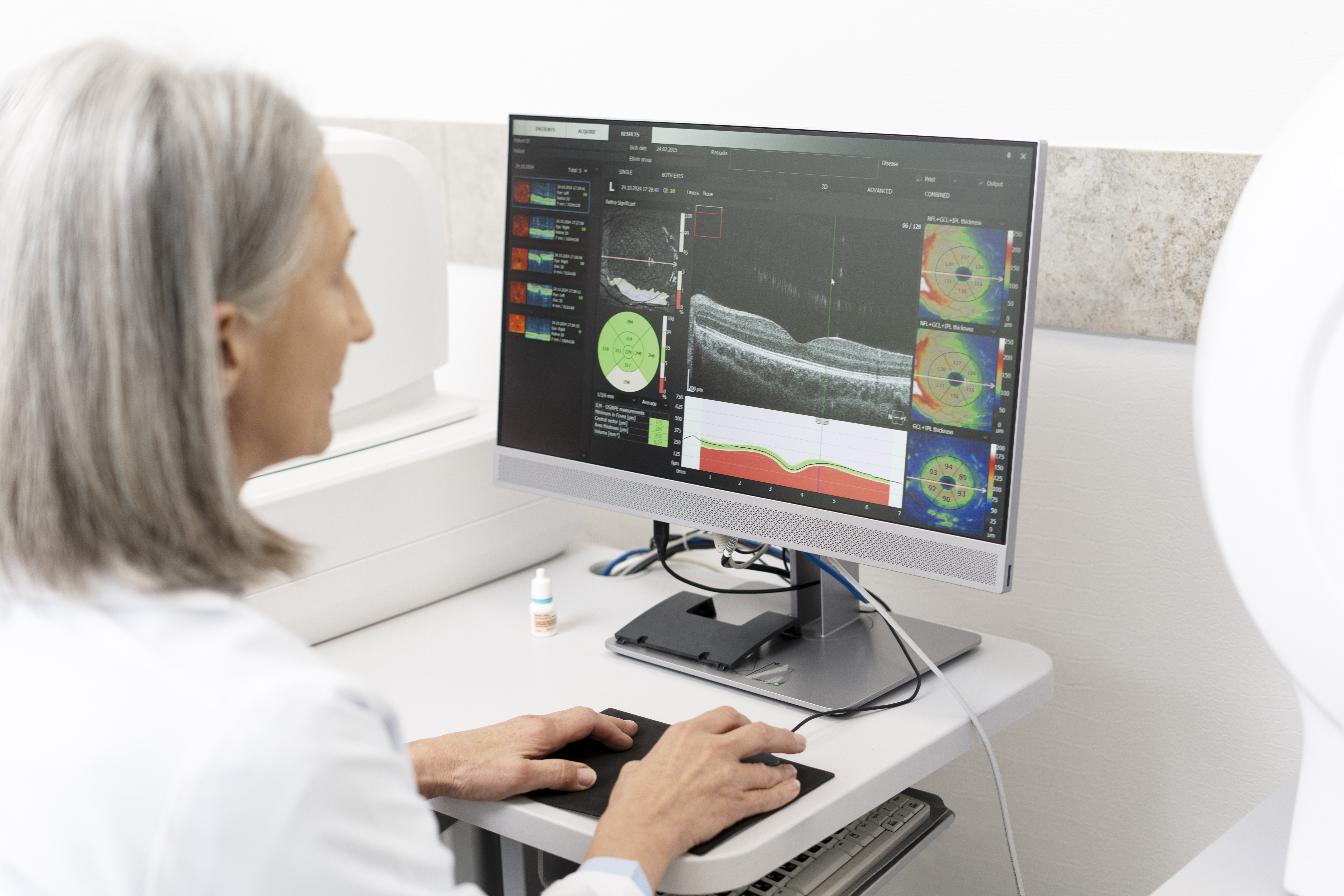
Role of Fluorescence Imaging with MolecuLight DX
The MolecuLight DX is an innovative tool for wound assessment. It uses fluorescence imaging to detect bacteria presence. This immediate feedback allows you to tailor treatment effectively.
Using this tool, you can guide cleaning and debridement more accurately. By focusing on areas with high bacterial load, you reduce infection risks. This precision can accelerate healing in stubborn wounds.
Benefits of Rapid Pathogen Testing
Detecting wound pathogens quickly is vital. Rapid pathogen testing identifies microbes faster than traditional methods. This speed means you can start the right treatment sooner.
This method allows for targeted antibiotic use, reducing unnecessary drug exposure. Patients benefit from faster recovery times, improving overall care quality.
Utilizing Triple-layer Allografts in Treatment
Triple-layer allografts are another advanced option. These allografts provide a scaffold for new tissue growth. Compliant with FDA regulations, they offer a regulated and safe option for healing.
These allografts are particularly useful in complex cases like pressure injuries. By supporting natural healing processes, they enhance recovery rates and improve patient outcomes.
When to Escalate Wound Care
Sometimes, wound healing requires more specialized intervention. Knowing when to escalate care is crucial for patient safety and recovery.
Indicators for Specialty Intervention
Certain signs suggest the need for specialized care. If a wound remains unresponsive after weeks of treatment, it may need advanced intervention. You should also consider escalation if there are signs of severe infection or underlying conditions like arterial disease.
Timely escalation can prevent complications. By partnering with specialists, you ensure the patient receives comprehensive care tailored to their needs.
Importance of Comprehensive Osteomyelitis Workup
In cases where bone infection is suspected, a thorough osteomyelitis workup is essential. This involves imaging and lab tests to confirm diagnosis. Early detection allows for prompt treatment, preventing further complications.
A comprehensive approach helps in managing complex cases. By addressing bone infection early, you can improve patient outcomes and reduce lengthy recovery times.
Enhancing Patient Outcomes with Wound Debridement
Wound debridement is a critical part of advanced care. By removing dead tissue, you create an optimal environment for healing. This procedure reduces infection risks and supports new tissue growth.
Incorporating regular debridement into treatment plans can significantly enhance recovery. Patients experience quicker healing and reduced discomfort, leading to improved satisfaction and outcomes.
In conclusion, understanding when and how to escalate care is key in managing stalled wounds. By leveraging advanced technologies and methods, you can ensure effective healing and patient satisfaction.
Seeing What Swabs Miss: How MolecuLight DX Elevates Wound Assessment Precision
Traditional swab tests often miss crucial bacterial details hidden deep within wounds. If you rely solely on those methods, you may overlook infection risks that slow healing. MolecuLight DX’s fluorescence imaging reveals bacterial fluorescence in real time, guiding precise wound assessment and treatment decisions. Keep reading to learn how this technology sharpens wound measurement and supports better outcomes for your patients.
Enhancing Wound Assessment Precision
Achieving accuracy in wound assessment is vital for effective healthcare. With innovative tools like MolecuLight DX, healthcare professionals can now reach precision levels previously thought unattainable.
The Role of MolecuLight DX
Imagine seeing beneath the surface of a wound without invasive procedures. MolecuLight DX provides that insight by using fluorescence imaging to reveal bacterial presence. This portable device helps you quickly identify problematic areas, ensuring you make informed decisions about patient care. Swab tests alone may miss harmful bacteria, delaying healing. With MolecuLight DX, you can detect these issues early, reducing the chances of complications. The tool’s real-time data allows for immediate responses, ensuring the right treatment path is followed. This approach not only optimizes patient care but also enhances your confidence in treatment decisions.
Fluorescence Imaging in Wound Care
Fluorescence imaging shines a light on hidden bacterial threats. By illuminating bacteria, this technique guides you in assessing wounds more thoroughly. Traditional methods might overlook biofilms, leading to prolonged recovery times. With fluorescence imaging, you pinpoint exact bacterial locations, allowing for targeted treatment. This method stands out in advanced wound care by providing visual evidence of infection, facilitating precise therapeutic actions. The images captured can be shared across your care team, ensuring everyone is on the same page regarding patient needs. As a result, care is more coordinated and effective.
Benefits of Accurate Wound Measurement
Precise wound measurement is more than just numbers; it’s about optimizing patient outcomes. MolecuLight DX offers a detailed view, enabling you to measure wounds accurately every time. This precision is crucial for tracking healing progress and adjusting treatments as needed. Inconsistent measurements can lead to inappropriate treatments, delaying recovery. By using this technology, you ensure that each assessment contributes to a comprehensive understanding of the wound’s status. This approach enhances treatment plans, promoting faster healing and reducing complications. Accurate data empowers you to make confident, informed decisions that benefit your patients.
Advanced Techniques in Infection Detection
Modern wound care requires advanced approaches to detect infections. MolecuLight DX offers groundbreaking techniques that elevate your practice’s ability to identify and address bacterial presence.
Bacterial Fluorescence and Bioburden Visualization
Understanding the bacterial load in wounds is crucial for effective care. MolecuLight DX enables you to visualize bacterial fluorescence, offering a clear picture of bioburden levels. This tool helps detect areas with high bacterial concentration, which can guide your treatment approach. Traditional methods may miss scattered bacteria, but this technology reveals them in vivid detail. By seeing what’s often invisible, you can address potential issues before they escalate. Sharing these visual insights with your team ensures everyone is aligned in the treatment approach, fostering a collaborative care environment.
Debridement Guidance and Therapy Selection
Choosing the right treatment depends on accurate information. MolecuLight DX aids in directing debridement efforts by highlighting areas needing attention. This guidance ensures that your interventions are as effective as possible. By targeting specific zones, you avoid unnecessary tissue removal, preserving healthy areas. Precision in debridement means that you can tailor therapy selection more accurately, enhancing patient outcomes. This approach minimizes risks, accelerates recovery, and ensures that each step taken is in the best interest of your patient.
Supporting Antibiotic Stewardship
Antibiotic stewardship is essential in today’s healthcare landscape. MolecuLight DX supports this by providing accurate bacterial insights. By identifying bacterial presence precisely, you can make informed decisions about antibiotic use. This reduces unnecessary prescriptions, combating antibiotic resistance. With this technology, you ensure that antibiotics are used only when truly needed, preserving their effectiveness. Your patients benefit from targeted treatments, reducing exposure to broad-spectrum antibiotics. This approach aligns with best practices in healthcare, promoting responsible use of medications.
Streamlining Wound Care Management
Efficient wound care management is key to patient satisfaction and recovery. MolecuLight DX streamlines various aspects of care, enhancing workflow and documentation processes.
Improving Wound Documentation
Accurate documentation is crucial for continuity of care. MolecuLight DX supports this by capturing detailed wound images, which you can easily integrate into patient records. These images provide a visual timeline of healing, making it easier to track progress. This tool simplifies documentation, ensuring that records are comprehensive and up-to-date. With clear visual evidence, you enhance communication with your team and patients, facilitating a shared understanding of treatment plans. Improved documentation leads to better patient outcomes and streamlined care processes.
Enhancing Wound Clinic Workflow
Clinic efficiency directly impacts patient experiences. MolecuLight DX enhances your workflow by enabling quick, precise assessments. This technology minimizes the time needed for wound evaluations, allowing you to focus on patient care. Faster assessments mean you can see more patients without compromising quality. By reducing bottlenecks in your clinic, you enhance the overall patient experience, fostering satisfaction and trust. Your clinic becomes a model of efficiency, leading to increased patient loyalty and improved outcomes.
Clinical Decision Support with MolecuLight DX
Making informed decisions is the cornerstone of effective care. MolecuLight DX provides the support you need for confident clinical decisions. With real-time bacterial visualization, you have the data necessary for precise treatment choices. This tool complements your expertise, offering insights that enhance your decision-making process. By incorporating this technology into your practice, you ensure that every decision is backed by solid evidence. This leads to improved patient outcomes, reduced complications, and a reputation for excellence in wound care.
Incorporating MolecuLight DX into your practice offers numerous benefits, from enhancing wound assessment precision to streamlining management. By leveraging this technology, you position yourself at the forefront of advanced wound care, ensuring the best possible outcomes for your patients.
Beyond Cultures: How Advanced Pathogen Detection Accelerates Wound Healing
Traditional wound infection diagnostics can take days, delaying critical treatment decisions. You know that every hour lost increases the risk of complications and graft failure. Advanced pathogen detection changes that timeline by providing rapid, precise results that guide targeted care and improve wound healing outcomes. In this post, you’ll learn how molecular wound testing and tools like MolecuLight DX support faster recovery and smarter antimicrobial stewardship. For more information, visit this article.
The Evolution of Wound Infection Diagnostics
Traditional methods in wound infection diagnostics have long relied on cultures, but advancements have paved the way for more precise solutions. Let’s explore how these innovations are shaping modern wound care.
From Cultures to Molecular Testing
Cultures have been the backbone of infection diagnostics, yet their limitations are clear. They require days to yield results, potentially delaying vital treatments. Enter molecular wound testing. This method, including PCR wound testing, provides results within hours. By identifying pathogens swiftly, you can initiate appropriate therapies sooner. For instance, MolecuLight DX allows for immediate visualization of wound biofilm, taking diagnostics beyond just numbers. It’s a game-changer in how you approach chronic wound management, especially in cases like diabetic foot ulcers.
Benefits of Rapid Wound Diagnostics
Rapid diagnostics offer more than just speed. They lead to better-targeted treatments, reducing unnecessary antibiotic use. Imagine the impact on antimicrobial stewardship: precise pathogen detection means you only use what’s necessary. Patients benefit too. Faster identification of bacterial load aids in quicker recovery and potentially fewer complications. In pressure injury care, this can dramatically alter outcomes. A study found that rapid diagnostics reduced treatment delays by 40%. This efficiency translates into improved care, especially for complex wounds like venous leg ulcers.
Role of Point-of-Care Imaging
Point-of-care imaging plays a crucial role in modern diagnostics. Technologies like fluorescence imaging allow for real-time assessment. With MolecuLight DX, you see bacterial presence directly, which guides targeted debridement. This visual insight ensures that treatments are precise, improving wound healing outcomes. Such tools are indispensable in environments where quick decision-making is vital. They transform how you monitor progress and adjust care plans, ensuring that each step is based on accurate, timely data.
Enhancing Wound Healing Outcomes
Next, we delve into how these diagnostic advancements enhance overall wound healing. The impact is far-reaching, from reducing therapy time to boosting graft success rates.
Reducing Time-to-Therapy
Time is of the essence in wound care. With advanced pathogen detection, therapy initiation is faster. Traditional methods might delay treatment decisions by several days. Now, molecular testing can slash this period to mere hours. In practice, this means patients receive timely interventions, drastically reducing the risk of complications. For healthcare professionals, it’s a relief knowing that each decision is backed by solid data, paving the way for proactive rather than reactive care.
Improving Graft Success Rates
Graft procedures demand precision. Any delay or misstep can lead to failure, something neither patients nor practitioners want. By using advanced diagnostics, you can better prepare for grafting procedures. Identifying the specific pathogens involved allows for tailored antimicrobial strategies, ensuring the wound environment is optimal for graft acceptance. This approach has been shown to increase success rates by up to 30%, a significant improvement that underscores the value of precise diagnostics in surgical settings.
Antimicrobial Stewardship in Wound Care
Antimicrobial stewardship is crucial, especially in today’s healthcare landscape. Rapid wound diagnostics support this by ensuring antibiotics are used appropriately. Instead of broad-spectrum treatments, you can target specific infections, reducing the risk of resistance. This precision helps preserve antibiotic efficacy, a critical component of chronic wound management. By being judicious with treatments, you not only ensure better patient outcomes but also contribute to the broader fight against antimicrobial resistance.
Specialty Wound Care as a Partner
Specialty Wound Care stands as a leader in this diagnostic revolution. Their programs and tools are designed to integrate seamlessly into your practice, enhancing care delivery.
Advanced Pathogen Detection Program Features
Specialty Wound Care offers comprehensive pathogen detection programs. These programs are tailored to meet the needs of modern healthcare facilities. They utilize cutting-edge technology, including the MolecuLight DX for real-time imaging and triple-layer allografts for wound healing. By partnering with Specialty Wound Care, you gain access to tools that streamline diagnostics and treatment planning, ensuring patients receive the highest standard of care efficiently.
Integrating MolecuLight DX in Practice
Integrating MolecuLight DX into your practice is straightforward. This tool offers clear advantages in daily wound assessment. It enhances your ability to visualize and document bacterial load, a crucial aspect of effective wound management. Training is provided to ensure seamless incorporation into your workflow, making it a valuable addition for any facility focused on precision-driven care. It’s not just about technology; it’s about empowering you to deliver better outcomes.
Requesting a Clinical Demo and Consultation
Interested in seeing these innovations in action? Specialty Wound Care offers clinical demos and consultations. This is your opportunity to experience firsthand how advanced diagnostics can transform your practice. By scheduling a demo, you can explore the potential benefits for your patients and streamline your wound care processes. It’s a step towards elevating the standard of care and ensuring you remain at the forefront of wound management excellence.
Accelerating Chronic Wound Healing With Triple-Layer Allografts
Chronic wounds resist healing despite standard care, costing time and resources you can’t afford to waste. Triple-layer allografts offer a biologic skin substitute designed to support angiogenesis, granulation tissue, and epithelialization in stubborn wounds like diabetic foot ulcers and pressure injuries. This post explains how integrating these allografts within a comprehensive wound care protocol can improve outcomes and streamline your clinical workflow. For more information, you can refer to this study.
Understanding Triple-Layer Allografts
To tackle chronic wounds effectively, understanding the science behind triple-layer allografts is crucial. These advanced solutions play a game-changing role in healing complex wounds and improving patient outcomes.
What is a Triple-Layer Allograft?
A triple-layer allograft is an advanced biologic skin substitute. It comprises three layers meticulously designed to support wound healing processes like angiogenesis, granulation tissue formation, and epithelialization. These layers derive from donated human tissues screened and processed to meet FDA regulations. The benefits of using an allograft include providing a scaffold for cellular growth and promoting faster recovery compared to traditional methods. It creates an environment conducive to healing by mimicking natural tissue functions.
Benefits of Allograft Applications
The application of allografts offers significant benefits: enhanced wound closure, reduced healing time, and lower infection risks. Studies show that wounds treated with allografts heal faster than those without. This improvement is due to the allograft’s ability to support cell migration and tissue regeneration. For patients, it means fewer complications and a quicker return to everyday life. For healthcare providers, it translates to more efficient use of resources and better patient satisfaction.
Indications for Chronic Wound Treatment
Triple-layer allografts are suitable for various chronic wounds, including diabetic foot ulcers, venous leg ulcers, and pressure injuries. These stubborn wounds require a comprehensive approach, and allografts are a critical component of treatment plans. By addressing the unique needs of each wound type, allografts enhance the body’s natural healing processes. Their use is particularly beneficial in cases where standard treatments have failed, offering a new hope for healing and recovery.
Protocols for Effective Wound Management
Integrating allografts into wound management protocols can make a significant impact. Comprehensive and tailored approaches ensure optimal healing and patient outcomes.
Comprehensive Wound Care Protocols
A well-rounded wound care protocol starts with a thorough assessment. This involves identifying wound type, size, and underlying conditions. Tailored plans should incorporate advanced therapies like triple-layer allografts to maximize healing. Each step of care, from cleaning to dressing, should aim to create an ideal environment for recovery. Implementing these protocols can lead to a 30% increase in healing rates, as supported by clinical studies.
Role of Debridement and Moisture Balance
Debridement is a critical step in preparing a wound for healing. Removing dead tissue reduces infection risks and promotes healthy tissue growth. Achieving proper moisture balance is equally vital. Wounds heal better in a moist environment, which prevents tissue drying and speeds up closure. This balance is a cornerstone of effective wound management, ensuring the allograft’s optimal performance.
Infection Control and Fluorescence Imaging
Infection control is paramount in wound care. Techniques like fluorescence imaging provide crucial insights into bacterial presence, guiding targeted treatments. Using tools like the MolecuLight DX, clinicians can visualize biofilms and ensure precise cleaning and treatment. This technology enhances decision-making and helps avoid complications, making it a valuable asset in wound management.
Advantages of Partnering with Specialty Wound Care
Choosing the right partner in wound care can significantly streamline processes and enhance treatment outcomes. Specialty Wound Care offers unique advantages in this area.
Support and Training for Clinicians
Specialty Wound Care provides extensive support and training for clinicians, empowering them with the latest knowledge and skills. This training ensures that healthcare providers are equipped to deliver top-notch care, utilizing cutting-edge technology and techniques. Continuous learning opportunities foster a culture of excellence and innovation in wound management.
Technology and Reimbursement Assistance
Access to state-of-the-art technology is a hallmark of Specialty Wound Care. Tools like the MolecuLight DX enhance wound analysis and treatment precision. Additionally, they offer reimbursement assistance, ensuring that clinicians can focus on patient care without financial hurdles. This support simplifies processes and improves the overall efficiency of wound care practices.
Scheduling and Consultation Opportunities
Specialty Wound Care offers flexible scheduling and consultation opportunities to meet the diverse needs of healthcare providers. By collaborating closely with your team, they ensure seamless integration of services and treatments. This partnership approach fosters a supportive environment for achieving exceptional patient outcomes, making Specialty Wound Care a valuable ally in advanced wound management.
A wound that doesn't heal is more than just a break in the skin; it's a complex clinical challenge that signals a disruption in the body's natural repair mechanisms. Chronic wounds, formally defined as those that fail to proceed through an orderly and timely reparative process, affect millions of people and represent a significant burden on healthcare systems.
Far from being a single entity, these persistent injuries arise from a multitude of causes, each requiring a distinct diagnostic and therapeutic strategy. Differentiating between the various types of chronic wounds is the foundational step toward effective management and successful outcomes. An accurate diagnosis prevents treatment delays, avoids therapies that could be harmful, and paves the way for advanced interventions that can accelerate healing. Most importantly, it helps prevent devastating complications like severe infection or amputation.
This guide provides a comprehensive roundup of the primary categories of chronic wounds, from common pressure injuries to complex arterial ulcers. We will explore the defining characteristics of each, offering clinicians, caregivers, and patients a clear roadmap for:
Identification: Pinpointing the specific wound type based on clinical features.
Management: Understanding common treatment approaches and when to seek specialty care.
Prevention: Implementing targeted strategies to reduce the risk of occurrence and recurrence.
By breaking down the key types of chronic wounds, this article offers actionable insights to improve diagnostic accuracy, streamline care, and ultimately support the healing journey.
1. Pressure Injuries (Pressure Ulcers/Bedsores)
Pressure injuries, often called pressure ulcers or bedsores, are localized damage to the skin and underlying soft tissue. They typically occur over bony prominences like the sacrum, heels, hips, and elbows. This type of chronic wound develops from intense and prolonged pressure, or pressure combined with shear or friction, which cuts off blood supply to the tissue, leading to cell death.
These injuries are a significant concern for individuals with limited mobility, such as those who are bed-bound, wheelchair users, or patients in long-term care facilities. The severity is categorized into stages, ranging from Stage 1 (non-blanchable redness on intact skin) to Stage 4 (full-thickness tissue loss with exposed bone, tendon, or muscle), and also includes unstageable and deep tissue pressure injuries.
Key Causes and Risk Factors
The primary cause is sustained pressure that exceeds capillary blood pressure, obstructing blood flow. Key risk factors include immobility, sensory impairment (like in spinal cord injuries), malnutrition, dehydration, and medical conditions affecting blood flow such as diabetes and vascular disease. Excessive moisture from incontinence can also weaken the skin, making it more susceptible to breakdown. Preventing skin breakdown is paramount in avoiding pressure injuries. A practical guide to effective perineal care highlights essential techniques for patient care.
Diagnosis and Treatment
Diagnosis begins with a thorough skin assessment, often using validated risk scales like the Braden or Norton scales to identify at-risk patients. Clinicians look for signs of tissue damage, such as changes in skin color, temperature, or firmness. Early detection is critical, and advanced diagnostic tools can play a role. Point-of-care fluorescence imaging, for example, helps clinicians detect and locate elevated bacterial presence in and around the wound, which can guide treatment and prevent infection. To explore how this technology aids in managing complex wounds, you can learn more about point-of-care wound imaging.
Treatment focuses on removing pressure from the affected area, keeping the wound clean and dressed, managing pain, and ensuring proper nutrition. Key strategies include:
Regular Repositioning: Turning and repositioning patients at least every two hours.
Support Surfaces: Using pressure-reducing mattresses, cushions, and other devices.
Wound Care: Selecting appropriate dressings to maintain a moist healing environment and debriding necrotic tissue when necessary.
Nutrition: Ensuring adequate intake of protein, calories, vitamins, and minerals to support tissue repair.
Referral to a specialty wound care clinic is recommended for non-healing Stage 2 wounds or any Stage 3 or 4 injuries, as they often require advanced therapies like debridement, specialized dressings, or negative pressure wound therapy.
2. Venous Leg Ulcers
Venous leg ulcers (VLUs) are the most common type of lower extremity ulcer, accounting for approximately 70% of all leg ulcers. They arise from chronic venous insufficiency (CVI), a condition where the veins in the legs, particularly the valves, fail to efficiently return blood to the heart. This leads to a buildup of pressure in the lower leg veins (venous hypertension), which causes fluid to leak into the surrounding tissues, leading to edema, inflammation, and eventual skin breakdown.
These wounds typically develop on the medial (inner) side of the lower leg, often just above the ankle, an area known as the "gaiter region." They are often shallow, have irregular borders, and may be accompanied by a significant amount of exudate (fluid drainage). The surrounding skin frequently shows signs of venous disease, such as varicose veins, hemosiderin staining (a brownish discoloration), and lipodermatosclerosis (hardening and tightening of the skin).
Key Causes and Risk Factors
The root cause of VLUs is sustained venous hypertension from CVI. Key risk factors that contribute to this condition include a history of deep vein thrombosis (DVT), varicose veins, obesity, multiple pregnancies, and occupations that require prolonged standing. Other contributing factors are advanced age, limited mobility, a family history of venous disease, and previous trauma to the leg.
Diagnosis and Treatment
Diagnosis is primarily clinical, based on the characteristic appearance and location of the ulcer along with a patient history consistent with CVI. An Ankle-Brachial Index (ABI) test is crucial to rule out significant arterial disease before initiating compression therapy, which is contraindicated in cases of severe arterial insufficiency. Doppler ultrasound studies can confirm venous reflux and identify incompetent veins.
The cornerstone of VLU treatment is managing the underlying venous hypertension. Key strategies include:
Compression Therapy: Applying sustained, graduated compression (usually 30-40 mmHg) via multi-layer bandages or stockings is the gold standard. This reduces edema and supports venous return.
Leg Elevation: Regularly elevating the legs above the level of the heart helps reduce swelling and venous pressure.
Wound Care: This involves cleaning the wound, managing exudate with appropriate absorptive dressings to maintain a moist environment, and debriding any non-viable tissue.
Exercise: Encouraging calf muscle pump exercises, such as walking or ankle flexions, improves circulation.
For complex, non-healing ulcers, a referral to a specialty wound care clinic is necessary. They may consider advanced treatments, including advanced biologic skin substitutes to promote healing. Explore how a dual-layer allograft can aid in healing venous leg ulcers. In some cases, surgical interventions like vein ablation may be required to correct the underlying venous reflux.
3. Diabetic Foot Ulcers
Diabetic foot ulcers (DFUs) are a serious complication of diabetes, developing from a combination of peripheral neuropathy (nerve damage causing loss of sensation), peripheral arterial disease (poor circulation), and impaired healing. These open sores, typically located on the bottom of the foot, occur in approximately 15% of individuals with diabetes and are a leading cause of non-traumatic lower limb amputations. Even minor cuts or blisters can progress into severe ulcers because the patient may not feel the initial injury.
Key Causes and Risk Factors
The primary contributors are neuropathy, which diminishes the protective sensation of pain, and peripheral arterial disease, which reduces blood flow needed for healing. Structural foot deformities like bunions or hammertoes can create pressure points, leading to callus formation and eventual breakdown. Poor glycemic control is a major risk factor, as high blood sugar levels impair immune function and slow the healing process. Other risks include a history of previous ulcers, improper footwear, and vision problems that prevent patients from noticing foot injuries.
Diagnosis and Treatment
Diagnosis involves a comprehensive foot examination, including testing for neuropathy (using a monofilament), assessing circulation (checking pulses and ankle-brachial index), and inspecting the ulcer. The wound is classified using systems like the Wagner Ulcer Classification Scale to determine its depth and the extent of infection or gangrene. Advanced imaging, such as point-of-care fluorescence imaging, can be crucial for detecting high bacterial loads that are not clinically apparent, helping to guide debridement and antimicrobial strategies to prevent limb-threatening infections.
Treatment is multifaceted and requires a coordinated approach to address the underlying causes. Key strategies include:
Offloading: Relieving pressure from the ulcer using total contact casts, walker boots, or specialized footwear.
Debridement: Regularly removing dead or infected tissue from the wound to promote healthy tissue growth.
Infection Control: Administering appropriate antibiotics for infected ulcers, guided by wound cultures.
Moist Wound Healing: Applying advanced dressings to create an optimal environment for tissue repair.
Glycemic Control: Working with the patient to maintain stable blood sugar levels.
For complex or non-healing ulcers, referral to a specialty wound care clinic is essential. Exploring advanced treatment options for diabetic foot ulcers can provide access to therapies like cellular and tissue-based products, which can significantly improve healing outcomes.
4. Arterial Leg Ulcers
Arterial leg ulcers, also known as ischemic ulcers, are wounds caused by inadequate blood supply to the lower limbs. These ulcers account for approximately 10-15% of all lower extremity wounds and develop when arteries become narrowed or blocked by conditions like peripheral arterial disease (PAD), often a result of atherosclerosis. The reduced arterial blood flow deprives tissues of essential oxygen and nutrients, leading to tissue death and the formation of a painful, difficult-to-heal wound.
These ulcers commonly appear on the toes, feet, heels, or ankles. They often have a characteristic "punched-out" look with well-defined edges, a pale or necrotic base, and minimal to no drainage. Patients frequently experience significant pain, especially at night or when the leg is elevated (rest pain), which is a sign of critical limb ischemia.
Key Causes and Risk Factors
The root cause of arterial ulcers is insufficient arterial circulation. The most significant risk factor is peripheral arterial disease (PAD), which is strongly associated with atherosclerosis. Other major contributors include smoking, diabetes, high blood pressure (hypertension), high cholesterol, advanced age, and a family history of vascular disease. Any condition that compromises blood flow to the extremities can increase the risk of developing these types of chronic wounds.
Diagnosis and Treatment
Diagnosis starts with a physical examination, noting the ulcer's appearance and location, along with assessing for signs of poor circulation like weak or absent pulses, cool skin temperature, and shiny, hairless skin on the leg. The primary diagnostic test is the Ankle-Brachial Index (ABI), a non-invasive measurement comparing blood pressure in the ankle to that in the arm. An ABI value below 0.9 indicates PAD and a high likelihood of an arterial ulcer.
Treatment is complex and requires a dual focus: managing the wound and restoring blood flow. The primary goal is revascularization, which is crucial for healing.
Vascular Assessment: Urgent referral to a vascular surgeon is essential to evaluate the extent of arterial blockage and determine options for restoring circulation, such as angioplasty, stenting, or bypass surgery.
Wound Care: The wound must be kept dry and protected from infection. Dressings are chosen to manage any minimal exudate without macerating the surrounding skin. Debridement of dry, stable eschar (necrotic tissue) is generally avoided until blood flow is improved.
Pain Management: Arterial ulcers are notoriously painful. Pain relief strategies are critical and may involve keeping the limb in a dependent (lowered) position to improve blood flow by gravity.
Risk Factor Modification: Addressing underlying causes is vital. This includes smoking cessation, managing blood sugar and blood pressure, and antiplatelet therapy.
Avoid Compression: Unlike venous ulcers, compression therapy is strictly contraindicated as it can further impede already compromised arterial blood flow.
Patients with non-healing arterial ulcers, severe rest pain, or signs of gangrene require immediate evaluation at a specialty wound care center or by a vascular specialist to prevent limb loss.
5. Mixed Etiology Ulcers (Venous-Arterial)
Mixed etiology ulcers are complex chronic wounds resulting from a combination of both venous insufficiency and peripheral arterial disease (PAD). These ulcers, which account for up to 15-20% of all lower extremity ulcers, present significant diagnostic and treatment challenges because the management strategies for venous and arterial disease are often contradictory. For example, compression therapy vital for venous ulcers can be harmful if significant arterial insufficiency is present.
These wounds often appear on the lower leg or foot, particularly in the "gaiter" area common for venous ulcers, but may have features of both wound types. They can be painful, slow to heal, and are frequently seen in elderly patients with comorbidities like diabetes, heart disease, and a history of smoking. The presence of both disease processes complicates healing by impairing both the delivery of oxygenated blood and the removal of waste products from the tissue.
Key Causes and Risk Factors
The dual causes are venous hypertension (from faulty vein valves) and inadequate arterial blood flow (from atherosclerosis). This combination creates a uniquely hostile environment for tissue health and wound healing. Risk factors are a blend of those for venous and arterial diseases, including advanced age, a history of smoking, diabetes, hypertension, high cholesterol, obesity, a history of deep vein thrombosis (DVT), and immobility.
Diagnosis and Treatment
Accurate diagnosis is crucial and begins with a comprehensive vascular assessment. The Ankle-Brachial Index (ABI) is a key initial test to quantify the degree of arterial compromise. A low ABI (typically <0.8) suggests significant PAD, while an ABI >1.3 may indicate calcified, non-compressible arteries, often seen in diabetics. A duplex ultrasound is essential to visualize blood flow, identify blockages in arteries, and assess venous reflux.
Treatment for these types of chronic wounds is a delicate balancing act that must address both underlying conditions. Management often requires a multidisciplinary team, including a vascular specialist.
Vascular Assessment and Intervention: The first priority is often to address the arterial disease. If severe PAD is present, revascularization procedures like angioplasty, stenting, or bypass surgery may be necessary to restore adequate blood flow before healing can occur.
Modified Compression Therapy: Once arterial flow is deemed sufficient (typically an ABI >0.6), modified or reduced compression can be carefully applied to manage the venous component. This must be done cautiously to avoid compromising arterial circulation.
Wound Care: Standard wound care principles apply, including debridement of non-viable tissue, managing exudate with appropriate dressings, and preventing or treating infection.
Lifestyle and Risk Factor Modification: Patients must receive counseling on smoking cessation, diet, exercise as tolerated, and managing underlying conditions like diabetes and hypertension.
Referral to a specialty wound care clinic and a vascular surgeon is strongly recommended for any patient suspected of having a mixed etiology ulcer due to the complexity of care and the high risk of limb-threatening complications if managed incorrectly.
6. Neuropathic Ulcers (Non-Diabetic)
Neuropathic ulcers are wounds resulting from a loss of protective sensation, which is not exclusively linked to diabetes. These ulcers arise from various neurological conditions that damage peripheral nerves, including spinal cord injuries, Hansen’s disease (leprosy), traumatic brain injuries, and hereditary sensory neuropathies. Much like their diabetic counterparts, these wounds develop due to repeated trauma or unnoticed pressure on an insensate area, typically on the feet or over bony prominences.
The inability to feel pain, pressure, or temperature means that minor injuries like blisters, cuts, or sustained pressure go undetected and untreated. Over time, this repetitive, unfelt trauma causes the skin and underlying tissues to break down, forming a chronic wound. Patients with conditions like spina bifida or those who have suffered severe nerve damage are particularly vulnerable to these types of chronic wounds.
Key Causes and Risk Factors
The fundamental cause is peripheral neuropathy leading to a loss of protective sensation. This sensory deficit is the primary risk factor, as it eliminates the body's natural warning system against injury. Key contributing factors include motor neuropathy, which can cause foot deformities like claw toes and high arches, creating abnormal pressure points. Autonomic neuropathy can lead to dry, cracked skin that is more susceptible to breakdown. Risk is heightened by ill-fitting footwear, foreign objects in shoes, and any form of repetitive mechanical stress on the insensate area.
Diagnosis and Treatment
Diagnosis is primarily clinical, based on a physical examination of the wound and a comprehensive neurological assessment to confirm sensory loss. A patient’s medical history is crucial for identifying the underlying neurological condition. Monofilament testing can quantify the degree of sensory loss. Imaging studies like X-rays may be used to check for underlying bone deformities or osteomyelitis, a common complication.
Effective treatment hinges on addressing the root cause: pressure and trauma. The core strategies are:
Aggressive Offloading: This is the most critical component. Total contact casting (TCC), removable cast walkers, or specialized footwear are used to redistribute pressure away from the ulcer.
Wound Bed Preparation: The wound must be kept clean, debrided of any non-viable tissue, and covered with appropriate dressings to foster a moist healing environment and prevent infection.
Infection Management: Neuropathic ulcers are highly prone to infection, which must be treated promptly and aggressively with antibiotics and, if necessary, surgical intervention.
Patient Education: Intensive education on daily self-inspection of feet and skin, proper footwear, and avoiding potential hazards is essential for both healing and prevention.
For complex, non-healing neuropathic ulcers, referral to a specialty wound care clinic is vital. Advanced treatments may include bio-engineered skin substitutes, surgical correction of foot deformities (e.g., tendon transfers), and hyperbaric oxygen therapy to support healing in compromised tissue.
7. Traumatic and Surgical Wounds
While many traumatic injuries and surgical incisions heal uneventfully, they can become chronic wounds when they fail to progress through the normal stages of healing within the expected timeframe, typically lasting longer than three months. This category includes a wide range of injuries, such as burns, deep lacerations, crush injuries, or surgical sites that break down (dehisce) or become infected.
The transition from an acute to a chronic state often occurs due to complicating factors that disrupt the delicate healing cascade. These issues can include persistent infection, poor blood supply to the wound area, significant tissue loss, or inadequate initial wound management. These types of chronic wounds are particularly challenging because they often involve complex damage to multiple tissue layers and may require extensive reconstructive efforts.
Key Causes and Risk Factors
The primary cause is a failure in the healing process following initial trauma or surgery. Key risk factors that predispose a wound to become chronic include severe contamination at the time of injury, the presence of foreign bodies, and extensive tissue damage that impairs vascularity. Systemic factors also play a huge role; patients with diabetes, peripheral vascular disease, compromised immune systems, or poor nutritional status are at a much higher risk. Surgical complications like hematomas, seromas, or high tension on the closure can also lead to wound breakdown and chronicity.
Diagnosis and Treatment
Diagnosis involves a detailed patient history, a physical examination of the wound to assess for signs of non-healing (e.g., persistent inflammation, lack of granulation tissue), and investigation of underlying causes. Cultures are often taken to identify infectious organisms, and imaging studies like X-rays or CT scans may be needed to check for foreign bodies or underlying bone infection (osteomyelitis). Advanced diagnostics, such as fluorescence imaging, can be invaluable for detecting bacterial presence in and around the wound, guiding targeted debridement and antimicrobial therapy to manage biofilm and prevent further complications.
Treatment is multifaceted and aims to address the root cause of the healing failure. Key strategies include:
Aggressive Debridement: Removing all non-viable (necrotic) tissue, foreign material, and bacterial burden is critical. This may be done surgically, enzymatically, or through other methods.
Infection Control: Systemic and topical antibiotics are used to manage infection based on culture results.
Advanced Wound Dressings: Dressings are selected to manage exudate, protect the wound bed, and create a moist environment conducive to healing.
Surgical Reconstruction: Wounds with significant tissue loss may require advanced procedures like skin grafts, flap reconstruction, or the use of tissue substitutes to achieve closure.
Optimizing Systemic Health: Addressing nutritional deficiencies, managing blood glucose levels, and improving circulation are essential for supporting the body's healing capacity.
Referral to a specialty wound care clinic or a plastic and reconstructive surgeon is often necessary for traumatic and surgical wounds that show signs of becoming chronic, especially in cases of dehiscence, exposed hardware, or extensive tissue loss.
8. Atypical and Inflammatory Ulcers
Atypical and inflammatory ulcers are a complex category of chronic wounds that arise from underlying systemic diseases rather than external factors like pressure or poor circulation. These conditions, which include pyoderma gangrenosum, vasculitis, and necrobiosis lipoidica, represent about 5-10% of all chronic wounds. They are frequently misdiagnosed as more common ulcer types, leading to incorrect treatments that can worsen the wound and delay proper care.
These ulcers often have unique clinical features, such as the rapidly progressing, purplish, undermined borders characteristic of pyoderma gangrenosum or the punched-out lesions seen in vasculitis. Because their root cause is an overactive or dysfunctional immune or inflammatory response, these wounds do not respond to standard wound care protocols and require a completely different management approach focused on treating the systemic disease.
Key Causes and Risk Factors
The primary cause of these ulcers is an underlying pathology that triggers inflammation and tissue destruction. For vasculitic ulcers, inflammation of blood vessels (vasculitis) restricts blood flow, causing skin to die. In pyoderma gangrenosum, a disorder of the immune system leads to painful, rapidly enlarging ulcers. Risk factors are directly linked to these systemic conditions, such as having inflammatory bowel disease (Crohn's disease, ulcerative colitis), rheumatoid arthritis, or other autoimmune disorders. A high index of suspicion is crucial for any wound that appears unusual, is extremely painful, or fails to heal despite appropriate standard care.
Diagnosis and Treatment
Diagnosis is often challenging and relies heavily on a thorough patient history, clinical presentation, and a definitive skin biopsy for histopathological examination. This biopsy is critical to distinguish these inflammatory ulcers from other types of chronic wounds, like infections or malignancies. Screening for associated systemic diseases through blood tests and consultations with specialists like dermatologists or rheumatologists is also a key part of the diagnostic process. Importantly, aggressive debridement must be avoided if pyoderma gangrenosum is suspected, as this can trigger a phenomenon called pathergy, causing the ulcer to expand rapidly.
Treatment is two-pronged, addressing both the systemic disease and the local wound. Management often requires systemic therapies to control the underlying inflammation, which may include:
Systemic Immunosuppression: Corticosteroids, biologics, or other immunosuppressive agents are often the primary treatment.
Gentle Wound Care: Protecting the wound with non-adherent, gentle dressings is essential to prevent further trauma.
Pain Management: These ulcers can be exceptionally painful, requiring a robust pain management plan.
Specialist Consultation: Collaboration with dermatology and rheumatology is vital for managing the underlying condition and adjusting treatments based on disease activity.
Referral to a specialty wound care clinic is recommended immediately if an atypical ulcer is suspected. These specialized centers can facilitate the necessary diagnostic workup, including biopsies, and coordinate care with other specialists to ensure the systemic cause is treated effectively.
Comparison of 8 Chronic Wound Types
Condition
Implementation complexity 🔄
Resource needs ⚡
Expected outcomes ⭐📊
Ideal use cases 💡
Key advantages ⭐
Pressure Injuries (Pressure Ulcers/Bedsores)
Low for prevention; high for advanced stages (surgical care)
Moderate–High: pressure-relief devices, staffing, possible surgery
📊 Good with prevention; prolonged healing and infection risk in advanced stages
Immobile/bed-bound patients, ICU, long-term care
Preventable, clear staging and established protocols
Venous Leg Ulcers
Moderate: sustained compression and monitoring
Moderate: compression systems, outpatient follow-up, possible venous procedures
📊 High healing rates with compression (>70%); recurrence common (30–40%)
Treatable with disease‑specific therapy; excellent healing potential once controlled
Integrating Advanced Diagnostics and Therapies for Better Outcomes
Navigating the complex landscape of chronic wounds, from pressure injuries to atypical inflammatory ulcers, requires more than just a foundational understanding. True mastery lies in recognizing the subtle yet critical differences between the various types of chronic wounds and deploying a sophisticated, multi-faceted approach to both diagnosis and treatment. As we've explored, the journey from initial assessment to complete healing is rarely linear. It is a dynamic process influenced by underlying comorbidities, patient adherence, and the specific etiology of the wound itself.
The core takeaway is this: accurate diagnosis is the cornerstone of effective wound care. A venous ulcer will not heal with treatments designed for an arterial insufficiency, and a diabetic foot ulcer requires a different offloading and glycemic control strategy than a post-surgical wound. Misidentifying the wound type or overlooking a mixed etiology can lead to stalled progress, increased patient suffering, and a higher risk of severe complications like infection, amputation, and sepsis. This is precisely why the initial assessment is so critical, demanding a thorough review of patient history, a comprehensive physical examination, and the use of appropriate diagnostic tests.
Embracing a Proactive, Technology-Driven Approach
The modern wound care paradigm is shifting away from reactive, trial-and-error methods toward a proactive, evidence-based model. This evolution is powered by technological advancements that provide clinicians with unprecedented insight into the wound environment.
Precision Diagnostics: Instead of waiting days for traditional culture results, rapid diagnostic testing can identify specific pathogens and their antibiotic sensitivities within hours. This allows for the immediate implementation of targeted antimicrobial therapy, preventing the overuse of broad-spectrum antibiotics and combating resistance.
Real-Time Visualization: Advanced fluorescence imaging tools like MolecuLight DX have revolutionized bedside care. By making high bacterial loads visible in real-time, clinicians can perform more precise, targeted debridement, ensuring all non-viable tissue is removed while preserving healthy granulation tissue. This technology also guides swab sampling for more accurate culture results.
Key Insight: Integrating these technologies creates a powerful feedback loop. Imaging guides treatment, rapid diagnostics confirm the microbial threat, and advanced therapies are applied with greater precision, dramatically accelerating the healing trajectory.
By combining these diagnostic insights with state-of-the-art therapies, such as cellular and tissue-based products (CTPs) like amniotic membrane allografts, we can create an optimal healing environment. These regenerative therapies provide a scaffold rich in growth factors, recruiting the body's own cells to rebuild damaged tissue more effectively. This synergistic model, which marries diagnostic clarity with therapeutic innovation, is the future of managing all types of chronic wounds.
Patient-Centered Care and Actionable Next Steps
Ultimately, the goal of understanding these wound types is to restore function and improve the patient's quality of life. Healing is not just a clinical outcome; it's a deeply personal journey. A holistic approach must therefore include robust patient education, empathetic communication, and a focus on the overall patient experience. Beyond the clinical aspects, patient experience is also vital for long-term care adherence. To ensure comprehensive patient care, it's beneficial to explore strategies for improving patient satisfaction scores.
For patients and caregivers, the most crucial next step is to seek specialized care at the first sign of a non-healing wound. Do not wait for complications to arise. For clinicians, the call to action is to embrace these advanced diagnostic and therapeutic tools and to build collaborative relationships with wound care specialists. By working together, we can ensure every patient receives an accurate diagnosis and a personalized, effective treatment plan, transforming the prognosis for even the most challenging chronic wounds.
Are you a clinician managing complex patients or a patient struggling with a non-healing wound? Partner with the experts at Specialty Wound Care to access the advanced diagnostics and evidence-based therapies needed to achieve superior outcomes. Visit Specialty Wound Care to learn how we bring specialized, comprehensive wound management directly to you.
Untreated wound pain can delay healing, increase stress, and diminish quality of life. Recognizing and managing this pain is crucial not just for comfort, but for overall recovery. A comprehensive approach to pain relief promotes faster healing and a better recuperation experience.
🧠 What Causes Pain in Chronic or Acute Wounds?
Pain from wounds may result from nociceptive pathways, nerve involvement, inflammation, ischemia, moisture imbalance, and trauma during dressing changes. When nerves are damaged, they can cause a sharp, shooting pain. Inflammation is typically the body’s response to injury, while ischemia refers to reduced blood flow causing pain. Explore diabetic foot ulcer resources for specific insights on nerve-related pain.
🩹 Dressing-Related Pain: What Can Be Done?
Managing pain during dressing changes is vital. Consider silicone-based dressings, atraumatic adhesives, and moisture-retentive materials. These options can reduce friction and discomfort. Techniques for gentle removal are equally important to prevent trauma. For detailed options, read about advanced wound dressing selections.
💊 Medication: Topical, Oral, and Injectable Options
Pain relief can include lidocaine gels for topical application, acetaminophen and NSAIDs for oral use, and nerve blocks or cautious opioid use for more severe pain. It’s important to assess each option with a healthcare provider to determine the best approach based on the wound type and patient needs. Explore our patient self-care guides for more insights.
🌿 Beyond Pills: Alternative Pain Relief Techniques
Alternative methods such as hydrotherapy, compression therapy, massage, and aromatherapy can offer pain relief without pharmaceutical intervention. Some patients find significant benefits from these tools in conjunction with traditional methods. See the latest innovations in wound care for more alternative strategies.
🧘 Mind-Body Tools That Help
Techniques such as deep breathing, guided imagery, meditation, and music therapy can be an integral part of holistic pain management. These approaches help reduce stress and anxiety, which can exacerbate pain perceptions.
👁️ Recognizing Pain When Patients Don’t Say It
Pain isn’t always verbalized. Caregivers should look for signs like withdrawal, facial expressions, moaning, and behavioral changes to assess discomfort in non-verbal patients. These cues can help tailor pain management strategies effectively. Wound care insights for specialty populations offer additional guidance.
🧑⚕️ What the Research Says
Recent studies have shown the effectiveness of multimodal pain management in chronic wound care settings. Combining various strategies often yields more effective results than relying on a single method. For real-life applications, view real patient healing stories to see how these methods are helping others.
📌 Pain Management Checklist
– When does it hurt most — dressing changes, walking, resting?
– Use topical relief when possible before procedures
– Choose non-adherent dressings for fragile skin
– Combine meds with mindfulness and movement
– Reassess pain every few days — don’t let it linger
📢 Call to Action
Pain relief isn’t just about comfort — it’s essential to healing. Explore how SpecialtyWoundCare.com supports your journey with expertise, empathy, and a commitment to pain-sensitive care.
Introduce the patient (use a fictionalized name): Sarah, a 55-year-old individual with a history of diabetes and long-term recovery from surgery. Sarah developed a diabetic foot ulcer due to poor circulation and nerve damage. Her emotional state was filled with anxiety and frustration as the wound refused to heal.
🔍 The Turning Point: Why They Sought Specialty Care
Sarah faced delays in wound healing, several misdiagnoses, and failed treatment attempts with previous providers. The wound became infected, worsening her symptoms and overall well-being. Desperate for a solution, Sarah sought out specialized wound care at SpecialtyWoundCare.com.
In the initial weeks, Sarah’s wound was assessed, and a personalized care plan was developed. Advanced wound dressings, offloading techniques, and nutritional adjustments were implemented. Through regular check-ups and monitoring, Sarah’s wound showed signs of improvement. By the fourth week, the wound began to heal from the inside out, reducing the risk of further complications.
Optional link: Read about advanced wound dressing options
🧪 What Made the Difference?
The key turning point in Sarah’s healing journey was the introduction of a moisture-balancing dressing that promoted a moist wound environment conducive to healing. This innovative approach accelerated the healing process significantly, leading to visible progress in just a matter of weeks.
Optional link: See the latest innovations in wound care
👩⚕️ The Team Behind the Scenes
Sarah benefited from a multidisciplinary team at SpecialtyWoundCare.com, including a registered nurse, podiatrist, wound specialist, dietitian, and social worker. This collaborative approach ensured that every aspect of Sarah’s care was addressed comprehensively, leading to more effective treatment outcomes.
Optional link: Learn how nutrition supports wound healing
📈 The Outcome: More Than Just Closure
Following weeks of specialized wound care, Sarah experienced a remarkable improvement in her quality of life. Not only did the wound close successfully, but Sarah also reported reduced pain, improved mobility, and a renewed sense of hope. The holistic approach taken by SpecialtyWoundCare.com restored not just Sarah’s physical health but her overall well-being.
Optional link: Tips for managing diabetic wound pain
📊 Patient Reflections
Sarah shared her fictionalized reflections, stating, “I never thought I would be able to walk without pain again. Thanks to the team at SpecialtyWoundCare.com, I am now able to enjoy activities like hiking pain-free, something I thought I had lost forever.”
📌 Key Lessons from This Case
– Early specialty care is crucial for preventing worsening outcomes.
– Maintaining moisture balance and proper offloading are critical for effective wound healing.
– Nutrition, movement, and emotional support play significant roles in the healing process.
– Healing is about restoring life and well-being, not just closing a wound.
Optional link: View real patient healing stories
📢 Call to Action
Every individual’s healing journey is unique. Discover how SpecialtyWoundCare.com provides personalized, compassionate care that addresses the holistic needs of each patient. Visit SpecialtyWoundCare.com to learn more about our comprehensive wound care services.
🔑 SUGGESTED SEO KEYWORDS
wound care case study, diabetic ulcer recovery, wound healing success, pressure ulcer treatment story, Specialty Wound Care Florida, chronic wound recovery, advanced wound therapy case
Meet Sarah, a 55-year-old with a history of diabetes. She developed a diabetic foot ulcer that persisted despite standard treatments, causing her immense distress and limiting her mobility.
🔍 The Turning Point: Why They Sought Specialty Care
Sarah experienced delays in healing her wound and faced misdiagnoses from previous providers. Frustrated, she turned to Specialty Wound Care for a fresh approach focused on her specific needs.
Over several weeks, Sarah underwent a comprehensive treatment plan at Specialty Wound Care. Dressings were meticulously chosen, offloading techniques were implemented, and nutritional adjustments were made to support her healing journey.
The key turning point for Sarah’s recovery was the adoption of a moisture-balancing dressing that significantly improved wound healing. This innovative approach marked a breakthrough in her treatment.
Sarah’s recovery was a collaborative effort involving a dedicated team at Specialty Wound Care — including nurses, podiatrists, wound specialists, dietitians, and social workers. Their combined expertise and support were instrumental in her progress.
Sarah’s life was transformed by the care she received at Specialty Wound Care. Not only did her wound heal, but she also experienced reduced pain, improved mobility, and a renewed sense of well-being.
“I couldn’t have imagined being pain-free again, but thanks to Specialty Wound Care, I can now enjoy long walks with my family every day,” Sarah joyfully shared.
Every healing journey is unique. Discover how SpecialtyWoundCare.com provides personalized and compassionate care to patients, focusing on holistic well-being.
Meet Sarah, a 55-year-old patient with Type 2 diabetes, who developed a stubborn diabetic foot ulcer after undergoing foot surgery. Sarah initially felt overwhelmed and anxious about the non-healing wound, unsure of how to manage the ongoing pain and discomfort.
🔍 The Turning Point: Why They Sought Specialty Care
Sarah endured months of ineffective treatments and misdiagnoses from various healthcare providers before deciding to seek specialized wound care. Frustrated by the lack of progress and worsening symptoms, she sought out experts in advanced wound care.
Week 1: Sarah’s wound was assessed by our wound care team, who recommended a tailored plan including regular dressing changes and offloading techniques to relieve pressure on the affected area.
Week 4: With the implementation of advanced wound dressings that promoted moisture balance, Sarah noticed a significant improvement in the healing process. Meanwhile, nutritional changes and emotional support played a crucial role in her overall well-being.
Optional link: Read about advanced wound dressing options
🧪 What Made the Difference?
The pivotal moment in Sarah’s healing journey came when the team introduced a cutting-edge moisture-balancing dressing that expedited the healing process. This innovative approach significantly accelerated the closure of the wound and minimized the risk of complications.
Optional link: See the latest innovations in wound care
👩⚕️ The Team Behind the Scenes
Sarah benefited from a holistic approach to her care, involving a multidisciplinary team of healthcare professionals. From registered nurses and podiatrists to wound specialists, dietitians, and social workers, each member played a vital role in coordinating her personalized treatment plan.
Optional link: Learn how nutrition supports wound healing
📈 The Outcome: More Than Just Closure
Following her treatment at SpecialtyWoundCare.com, Sarah experienced a remarkable improvement in her quality of life. Not only did the wound heal successfully, but she also noticed a significant reduction in pain and an enhancement in her mobility. The impact of the treatment extended far beyond just wound closure.
Optional link: Tips for managing diabetic wound pain
📊 Patient Reflections
Sarah shared, “I never imagined I would regain my mobility and live pain-free. Thanks to the compassionate care and expertise at SpecialtyWoundCare.com, I can now enjoy activities that were once a struggle for me.”
📌 Key Lessons from This Case
– Early intervention with specialty care is crucial for positive outcomes
– Maintaining moisture balance and proper offloading techniques are essential for wound healing
– Nutrition, emotional support, and collaborative care contribute significantly to the healing process
– Healing involves restoring overall well-being, not just the physical wound
Optional link: View real patient healing stories
📢 Call to Action
Every patient’s healing journey is unique. At SpecialtyWoundCare.com, we are dedicated to providing personalized and comprehensive care that addresses the individual needs of each patient. Discover how our expertise and compassionate approach can make a difference in your wound care journey.
Meet Jane, a 58-year-old woman with diabetes who struggled with a non-healing foot ulcer for six months. The wound developed following a minor injury that turned into a persistent problem. Jane, once active and independent, now faced the emotional turmoil of chronic pain and limited mobility.
Optional link: Explore diabetic foot ulcer resources
🔍 The Turning Point: Why They Sought Specialty Care
Frustrated by delayed healing and unsuccessful treatments, Jane turned to SpecialtyWoundCare.com out of desperation. Her previous healthcare providers lacked expertise in wound care, resulting in misdiagnoses and ineffective interventions. Seeking specialized care was her last hope for recovery.
Optional link: Explore our patient self-care guides
📆 Week-by-Week Healing Timeline
Week 1-2: Upon evaluation, our wound care team identified Jane’s wound type and initiated a treatment plan. Specialized dressings were applied to promote healing, and offloading strategies were implemented to reduce pressure on the affected area.
Week 3-4: Regular monitoring and adjustments were made according to Jane’s response to treatment. Lab tests revealed nutritional deficiencies, prompting dietary changes to support wound healing.
Week 5-6: Imaging studies showed the progress of tissue regeneration. Emotional support and encouragement played a crucial role in Jane’s motivation and resilience throughout the healing process.
Optional link: Read about advanced wound dressing options
🧪 What Made the Difference?
The pivotal moment in Jane’s recovery came with the introduction of a moisture-balancing dressing. This innovative approach optimized the wound environment, facilitating faster healing and preventing complications. The decision to switch to this advanced dressing significantly accelerated the healing progress.
Optional link: See the latest innovations in wound care
👩⚕️ The Team Behind the Scenes
A multidisciplinary team, comprising wound care specialists, podiatrists, dietitians, and social workers, collaborated to address Jane’s comprehensive needs. Their coordinated efforts ensured a holistic approach to her care, focusing not only on wound management but also on her overall well-being.
Optional link: Learn how nutrition supports wound healing
📈 The Outcome: More Than Just Closure
Beyond wound closure, Jane experienced a remarkable improvement in her quality of life. The reduction in pain and the restoration of mobility empowered her to resume her daily activities with newfound confidence. The impact of specialty wound care extended far beyond physical healing, enhancing Jane’s overall well-being.
Optional link: Tips for managing diabetic wound pain
📊 Patient Reflections
Reflecting on her journey, Jane shared, “I didn’t think I’d walk pain-free again — but now I’m hiking every Sunday. The team at SpecialtyWoundCare.com truly changed my life.”
Optional link: View real patient healing stories
📌 Key Lessons from This Case
1. Early specialty care prevents worsening outcomes
2. Moisture balance and offloading are critical
3. Nutrition, movement, and emotional support matter
4. Healing is about restoring life — not just skin
Optional link: View real patient healing stories
📢 Call to Action
Every wound care journey is unique, requiring personalized attention and expertise. Experience the difference with SpecialtyWoundCare.com, where we prioritize holistic care to achieve optimal outcomes for our patients.